tree in bud opacities pneumonia
Distal pulmonary vasculature More specifically the pattern can be manifest becaus. Multiple causes for tree-in-bud TIB opacities have been reported.
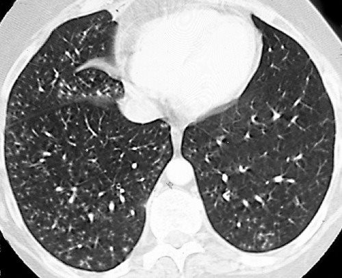
There are tree-in-bud opacities scattered throughout both lungs.

. The differential for this finding includes malignant and inflammatory etiologies either infectious or sterile. Pneumonia due to respiratory syncytial virus in a 23-year-old man with leukemia. Adjacent bronchial wall thickening is also frequently depicted.
Ad Do you have pneumonia. And tree-in-bud branching opacities detected throughout both lung fields after aspiration. 1 It is important for clinicians to remember that this pattern has an extensive.
Related
Tree-In-Bud Pattern A lymphoid interstitial infiltrate in the walls of the small airways follicular bronchiolitis may cause small centrilobular nodules and the tree-in-bud pattern Fig. Note the scattered lung nodules surrounded by halos of ground-glass attenuation. We here describe an unusual cause of TIB during the COVID-19 pandemic.
Associated focal ground-glass and consolidative opacities may be visualized although this should not the predominant feature. Studies have reported that pulmonary TB accounts for only 28 of the cause of tree-in-bud opacities as opposed to pulmonary apical granulomas and fibrosis being more suspicious of. A chest radiograph showed bilateral nodular opacities with a left lower lobar consolidative opacity Fig 1A 1B.
Tree-in-bud TIB appearance in computed tomography CT chest is most commonly a manifestation of infection. Interstitial pneumonia Parenchymal infection. Learn everything you need to know about pneumonia types.
TIB opacities represent a normally invisible branches of the bronchiole tree 1 mm in diameter that are severely impacted with mucous pus or fluid with resultant dilatation and budding of the terminal bronchioles 2 mm in diameter1 photo. A tree-in-bud pattern of centrilobular nodules from metastatic disease occurs by two mechanisms. 1 direct filling of the centrilobular arteries by tumor emboli and 2 fibrocellular intimal hyperplasia due to carcinomatous endarteritis.
The tree-in-bud pattern can be an early sign of disease Fig 10 15. A Thin-section CT scan of the right lung shows centrilobular ground-glass opacities in addition to nodules and tree-in-bud opacities arrow. More extensive lympho - cytic infiltrations may be associated with lymphoid interstitial pneumonia LIP with ground-.
Find out the 6 common types. Logic findings leads to a more accurate diagnosis. Aspiration Pneumonia and Tree in Bud Sign 87 year old male with history of cough and suspicion of aspiration shows barium aspiration into the.
Forms include secondary bacterial pneumonia mixed bacterial and viral pneumonia or primary influenza pneumonia. The purpose of this study was to determine the relative frequency of causes of TIB opacities and identify patterns of disease associated with TIB opacities. Since the initial report of endobronchial spread of pulmonary tuberculosis the tree-in-bud sign has been reported in a wide variety of health conditions including infectious diseases aspiration pneumonia congenital disorders idiopathic disorders inhalation immunologic disorders connective disorders 23456 and central lung cancer involving the.
Seasonal influenza in adults. Note the scattered lung nodules surrounded by. There is no lower lobe predominance as the distribution is quite diffuse.
Simply put the tree-in-bud pattern can be seen with two main sites of disease 3. However gram staining and cultures were negative. 2 However the classic cause of tree-in-bud is Mycobacterium tuberculosis especially when it is active and contagious and associated with cavitary lesions.
It is most commonly associated with infectious diseases affecting the bronchioles1 OP resulting in a tree in bud pattern has been previously suggested2 However a clear radiological-pathological correlation of OP filling the bronchioles resulting in a tree in bud pattern has to the best of our knowledge not yet been clearly demonstrated. Classically bronchiolitis appears as a region of centrilobular nodularity often in a tree-in-bud pattern. Thin-section CT scan shows peripheral poorly defined centrilobular nodules and tree-in-bud opacities bilaterally.
A young male patient who had a history of fever cough and respiratory distress presented in the emergency department. A young male patient who had a history of fever cough and respiratory distress presented in the emergency department. Tree-in-bud refers to a pattern seen on thin-section chest CT in which centrilobular bronchial dilatation and filling by mucus pus or fluid resembles a budding tree.
The low Gaffky score may have been secondary to the detection of mycobacteria other than M tuberculosis and findings related to food debris digestive juices and saliva. Tree-in-bud TIB opacities are a common imaging finding on thoracic CT scan. Malignancy can be associated with.
Tree-in-bud TIB appearance in computed tomography CT chest is most commonly a manifestation of infection. Distal airways more common 2. The tree-in-bud sign is a nonspecific imaging finding that implies impaction within bronchioles the smallest airway passages in the lung.
1012 Poorly defined centrilobular nodules associated with branching linear and nodular opacities ie tree-in-bud sign are the typical HRCT findings of infective bronchiolitis frequently. These small clustered branching and nodular opacities represent terminal airway mucous impaction with adjacent peribronchiolar inflammation. The imaging manifestations of small airways disease on high-resolution computed tomography may be direct or indirect signs of small airway involvement and include centrilobular nodules and branching nodular tree-in-bud opacities or the demonstration of mosaic attenuation that is typically exaggerated on expiratory computed.
3 Aspiration is also a common cause of the tree-in-bud formation. The patient underwent CT scanning of the chest which showed areas of nodular infiltration in the lower lobes with tree. Pneumonia due to respiratory syncytial virus in a 23-year-old man with leukemia.
There are two major pathologic patterns of viral pneumonia. However to our knowledge the relative frequencies of the causes have not been evaluated. Thin-section CT scan shows peripheral poorly defined centrilobular nodules and tree-in-bud opacities bilaterally.
Usually somewhat nodular in appearance the tree-in-bud pattern is generally most pronounced in the lung periphery and associated with abnormalities of the larger airways. There is no associated bronchiectasis bronchial wall thickening consolidation cavitation or lymphadenopathy. Mycobacterium avium complex is the most common cause in most series.
As in this case renal cell carcinoma is one of the most common malignancies that may produce this vascular cause of tree-in-bud pattern. Although initially described in 1993 as a thin-section chest CT finding in active tuberculosis TIB opacities are by. Patients with normal standard physiological pulmonary tests have been shown to have mosaic perfusion and air trapping on HRCT suggestive of bronchiolitis obliterans and a pattern of branching linear opacities like a tree in bud appearance suggestive of bronchiectasis with mucoid secretions.
4 His clinical course indicated a diagnosis of suspected DAB from centrilobular micronodules and treeinbud branching opacities detected throughout both lung fields. In the acute phase bacterial pneumonia manifests in the form of segmental or lobar consolidation Fig 2 possibly with cavitation and related hilar and mediastinal adenopathies.

Tree In Bud Caused By Haemophilus Influenzae Radiology Case Radiopaedia Org

Pdf Tree In Bud Semantic Scholar
View Of Tree In Bud The Southwest Respiratory And Critical Care Chronicles

Chest Ct With Multifocal Tree In Bud Opacities Diffuse Bronchiectasis Download Scientific Diagram
Tree In Bud Pattern Pulmonary Tb Eurorad
2
2
2
2
View Of Tree In Bud The Southwest Respiratory And Critical Care Chronicles

Tree In Bud Sign Lung Radiology Reference Article Radiopaedia Org

Tree In Bud Caused By Haemophilus Influenzae Radiology Case Radiopaedia Org

Hrct Scan Of The Chest Showing Diffuse Micronodules And Tree In Bud Download Scientific Diagram

Co Rads 2 With Tree In Bud Sign A 27 Year Old Male Attended The Download Scientific Diagram

Tree In Bud Sign And Bronchiectasis Radiology Case Radiopaedia Org

Tree In Bud Pattern Pulmonary Tb Eurorad

Pdf Tree In Bud

Ct Scan Of Chest Revealing Scattered Tree In Bud Opacities In Both Download Scientific Diagram
2